The properties of skins and parchment
General properties of skins
It is simpler to determine the damage suffered by parchment and analyze its status if you know its properties. Skins have a layered structure and their properties differ by layer.
Fundamentally, all mammal skins have a similar fibrous structure.
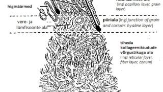
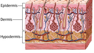
Three layers with different properties will be clearly visible in the cross-section of a mammal’s skin: the epidermis, dermis and the subcutaneous tissue (hypodermis). These simplified diagrams (ill 1-4) will help explain the structure of the skin.
On average, the raw skin used to manufacture parchment, initially contains 64% water, 33% protein, 2% fats, 0.5% mineral salts and 0.5% other substances (pigments, etc.).
In turn, the proteins contain 0.3% elastin, 29% collagen, 2% keratin and 1% non-structure proteins (albumin, globulin). The three visually discernible layers of skin contain different levels of these substances. The most collagen-rich part of the skin is the dermis, the most keratin-rich is the epidermis, and the most fat-rich is the subcutaneous tissue .
In the skin layers, the location of the blood and lymphatic vessels, as well as the size of the blood vessels, also differs (ill 3)

Layers of the skin
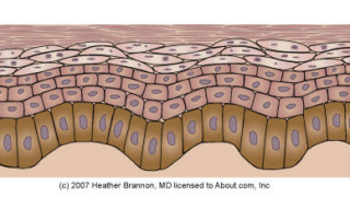
The epidermis is the outer surface of the skin and is basically comprised of protein keratin, which protects the lower layers of skin from the impact of the external environment. This is also the area where the hair follicles, sweat and sebaceous glands are found. The epidermis is connected to the lower layer of the skin by blood vessels, skin tissue and hairs. This skin layer can be dozens of times thicker in one area of an animal than another. For instance, the thickness of the epidermis around the eyes can be 0.05 mm and up to 1.5mm around the limbs. The epidermis is comprised of six layers that are vital to the functioning of a living organism (ill 4).
The Latin names, starting from the bottom are: stratum basale, stratum spinosum, stratum granulosum, stratum licidum, stratum corneum. The top layer, the stratum corneum or horny layer is an area with dead cells that contains solid keratin. The creation of new cells occurs in the bottom layer (stratum basale). The necessary nutrients reach this layer through the blood vessels in the dermis. The life span of the cells created in this layer is approximately two weeks.
The dermis is an area with a dense network of collagen fibers. The fibrous structure of this area is comprised of tightly intertwined bundles of collagen fibers (ill 1, 2). Depending on the species of animal, its diet and age, the thickness of the dermis may be between 1 and 3 mm.
The dermis consists of two visually discernible layers: the grain layer and the reticular layer
In addition to these discernible layers, the dermis also has three types of tissue with various properties and functions. These are dispersed in the dermis, not by layer, but throughout the entire network of fibers. These include collagen, which determines the strength of the skin, elastin that determines the flexibility and elasticity, and the tissue that contains the proteins to create the network of tissues.
The grain layer of the dermis has great absorption capacity, and based thereon, is chemically unstable. Chemically, the area separating the two layers (junction of grain and corium: hyaline layer) is loosely connected to the surrounding layers, thereby making it easy to physically remove the epidermis with its hairs and the network of blood and lymph vessels when processing the skin. This process is called skin splitting.
The fibers and bundles of fibers are separated from each other by an inter-fiber material. The thickness of the fiber network, diameter and orientation of the fibers are of determinative importance when it comes to the strength and flexibility of the skin. On top of the dermis, the diameter of the fibers is smaller and their orientation as related to the outer layer of skin differs from that of the lower layer of the dermis. The fibrous structure is clearly visible on the photos made by the scanning electron microscope in the Materials Engineering Research Centre at Tallinn University of Technology (ill 5, 6).
Basically, the subcutaneous layer is comprised of fat. This layer is unevenly spread over the entire skin of the animal and is structurally weak. When the skin is processed, this layer is physically removed.
Properties of parchment
When manufacturing parchment, the raw skin is processed and burnished on both sides so that the parchment is basically comprised of a dermis with a dense network of collagen fibers with a collagen content of almost 95%. The properties of the parchment are largely dependent on which part of the dermis is used to make the parchment.
The surface of the parchment will be shiner and physically stronger if, during the manufacture of the parchment, more of the underside is ground off, and the top layer, which contains finer collagen fibers, is left intact. However, the parchment will have a soft, velvety finish if more of the top layer of the dermis is ground off and the area with a softer, coarser fibrous structure on the underside of the skin is left intact.
When determining the status and damage suffered by the parchment, it is important to know that the structure of the parchment is hierarchical and contains collagen. Collagen is a protein, a high molecular compound, the monomers of which are various amino acids that are joined by peptide bonds.
The fibrous structure of parchment is organized as fiber bundles, fibrils (with a diameter of 250-500 nm) that have developed from microfibrils with the help of hydrogen bonds, covalent bonds and salts (ill 5). Collagen microfibrils, as collagen molecules, are exceptionally long (ca 300 nm) as compared to their cross-section (1.4 nm) and ca 1,000 units are contained in one molecular chain.
Tropocollagen molecules are the primary elements of parchment’s fibrous structure (ill 1). As a result of the specific combination of three polypeptide chains (i.e. amino acids connected to each other with the help of peptide bonds), collagen as a molecular structure of protein develops into a twisted left-handed spiral, an α-helix. When these twisted molecules intertwine, they form tropocollagen that is structured as a triple spiral right-handed super helix. This hierarchal structure gives the collagen molecules great stability and strength.
The triple spiral tropocollagen structure is the main unit of parchment’s hierarchical fibrous structure. The main forces holding the main triple helixes together are the hydrogen bonds between the triple helix and the covalent cross links with adjacent amino acid molecules (ill 2).
The long collagen molecules have overlapped ¼ of their length (i.e. 67nm) as so-called ‘transfers’. The microfibrils are located in the fibrils as staggered regions) (d). These repeated transfer areas with fixed frequency are clearly visible as dark stripes when examined under an electron microscope. (ill 1,2).
In summary, the hierarchical composition of the fibrous structure of the parchment means that microfibrils are formed from the tropocollagen molecules. Fibril is formed from the microfibrils and, in turn, collagen fibers and bundles are formed from the fibrils. (ill 2).
The structural composition and chemical properties of the parchment are closely linked.
Thanks to the hierarchical composition of the parchment, the collagen fibers can be very resistant to aging. The strength of the collagen molecules is derived from the hydrogen bonds between the chains and the cross-links in the molecules and microfibrils. This structural stability is preserved if no reasons exist for the triple helix structure to break down. There can be many reasons why the structure breaks down (E. Keedus, Pärgamendikollektsiooni konserveerimine ja säilitamine Ajalooarhiivis. MA thesis. Tartu 2006).
Studies conducted at the collagen fiber level confirm the instability of the parchment properties in relationship to the impact of the external environment. Excessive moisture, heat, radiation, environmental pollution, and other chemical influences contribute to the breakdown of the triple helix structure and hierarchal composition.
For instance, water and heat can cause the collagen fibers to swell and shorten, and the hydrogen bonds holding the three peptide chains together can break. Thus, the freed peptide chains form another, less ordered, structure such as gelatin. The gelatin formed from collagen fibers is the suspension of the parts of a polypeptide chain of different lengths in water, which wraps around the fibers, thereby making the fibers of the fibrous texture less distinctive. This change in the parchment is clearly visible as a distinctive glass-like layer on the surface, which is direct evidence of the gelatinization or degrading of the parchment.
The deterioration of the condition of historical parchment documents alludes primarily to changes in the chemical properties of the parchment, which are accompanied by irreversible structural changes. Less research has been done on how the non-collagen components impact the stability of the properties of historical parchment. For instance, lipids, which are non-polar biomolecules of the skin with an esteric structure that are part of the skin, and do not dissolve in water.
Lipids include fats, oils, waxes, etc. The fact that lipids do not dissolve in water makes their total elimination during the manufacture of parchment, which takes place in a damp environment or water, problematic. When manufacturing parchment, after the coat is removed the fats are usually removed physically, by scraping. However, the removal of the fats is hampered if the entire grain layer of the dermis is not removed along with the hair follicles that extend through this layer and form deep inclined depressions (ill 3,3a). It is difficult to remove the fats, as well as other residue of parchment manufacturing, from these depressions. The degradation or gelatinization of the parchment’s properties often starts from these hair follicles.
Studies conducted in recent years confirm the direct impact of the oxidation of lipids on parchment degradation (C. Ghioni, jt. Evidence of a Distinct Lipid Fraction in Historical Parchments: A Potential Role in Degradation?, 24.03.2016; A. Mozir jt., An Oxidative Degradation of Parchment and its Non-destructive Characterization and Dating, Appl. Phys. A 104 (2011), pp. 211–217).
The results of the studies have shown that the greater the lipid content in historical parchments, the greater the degree of degradation. The lipid content of a parchment may have been caused by how the manufacture of the parchment was conducted, by the bacterial impact of the environment, or the subsequent handling of the documents.
The fact that the gelatinization may be related to the hair follicles in the grain layer also significantly impacts the appearance of the parchment. In these cases, the parchment in the follicle area is significantly yellower and the resulting patterns of hair follicles is clearly discernible.
If the parchment was manufactured so that part of grain layer was left intact, the animal species can be identified based on the pattern of hair follicles. When identifying the animal species based on the hair follicles, the use of the following methodological materials is recommended: Helpfile — Parchment Assessment Report / IDAP March 2009; Conservation of Leather and Related Materials, M. Kite, R. Thomson. Oxford, 2006.
In the case of parchment, it is more difficult to determine the animal species based on the pattern of the hair follicles and this often requires the help of an expert. This is because the grain layer with the hair follicles is usually ground off when manufacturing parchment and the stretching usually distorts the pattern of the follicles on the surface.
In illustrations 4, 4a and 5, 5a, we see a comparison of the hair follicle patterns of one and same species of animal that can occur on tanned leather and degraded historical parchment (Codex Sinaticus, 24.03.2016 ).
All mammals have the following skin characteristics, which are thereby also visible on parchment:
- Specific pattern of hair follicles
- Diameter of collagen fibers
- Spatial placement of helixes
- Thickness of the skin
- Different structure and thickness of the three skin layers (epidermis, dermis, subcutaneous tissue).
These characteristics determine the physical and chemical properties of the skin. Thus, thanks to its structure, goatskin is physically stronger than sheepskin.
There are also large differences between old and young animals of the same species of mammal.
The skin of an animal is not consistent throughout. The structure of the skin in animal’s neck area differs from the area along its stomach, as does its density, chemical composition, general thickness and thickness of the three skin layers (epidermis, dermis, subcutaneous tissue).
It is also important to know that on the surface of a single mammal’s skin, the fibrous structure as well and the orientation of the fibers may differ in places, ill.5 (Parchment. The Physical and Chemical Characteristics of Parchment and the Materials Used in Its Conservation, B.H. Haines, 1999).
The stretching and shrinkage of the skin occurs more readily in the different orientation directions of the skin fibers as shown on ill.10. This is important to know in regard to the manufacture of leather items and the preservation of parchment. Due to the similar orientation of the fibers in the different directions in the middle of the skin (Section A), the skin is almost equally stretchable in different directions. However, if the parchment sheet has been cut from the skin in such a way that the document included areas A, B, C and D with their differing orientations, there would be a greater probability that the surface of the parchment would deform if the environmental conditions (humidity, temperature) changed.
Summary
In summary, it is important to know the following about the properties of skins and parchment:
- When determining the damage suffered by parchment, it is very useful to be familiar with the properties of the skin.
- In order to manufacture high-quality parchment, the skin of younger animals should be used.
Thin, elastic, and durable parchment and vellum is made from the skins of unborn or newly born lambs and calves. - High-quality, durable parchment is physically processed on both sides and is comprised mainly of the dermis with ultra-fine blood vessels from the most collagen-rich (more than 95% collagen) areas with a dense reticular layer.
- The sheets of parchment that have been cut out so as to eliminate the areas of the limbs (ill 10, D-area) and the edges of the stomach (B-area) are less likely to deform in the future.
The damage to and degrading of the parchment is irreversible if the hierarchic structure of the parchment has been destroyed.


